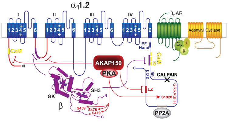
FIG. 6.
The Cav1.2-AKAP150-PKA complex. Blue, α11.2; magenta, β2 and its interactions with α11.2; yellow, CaM binding sites on α11.2 [there is one binding site in the NH2 terminus and three binding sites in tandem in the COOH terminus; the latter region also interacts with β subunits (magenta bracket and segment)]; red, AKAP79/150 binding sites (LZ, brackets, and segments), PKA, and PKA phosphorylation sites on α1 and β2 (arrows); gray, PP2A binding site; black X, calpain cleavage region; green, β2 AR; yellow-green, heterotrimeric G protein complex; yellow-orange, adenylyl cyclase.

