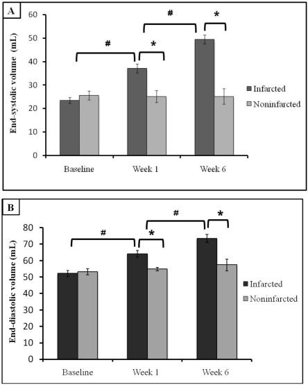
Figure 1.
Left ventricular remodeling before (baseline) and 1 and 6 wk after acute MI in swine. (A) End-systolic and (B) and end-diastolic volumes (mean ± SEM) are significantly increased when compared with values at baseline and from control (noninfarcted) animals. *, P < 0.05 between values for infarcted and noninfarcted animals at the same time point; #, P < 0.05 between values from infarcted animals at successive time points.

