
Figure 3.
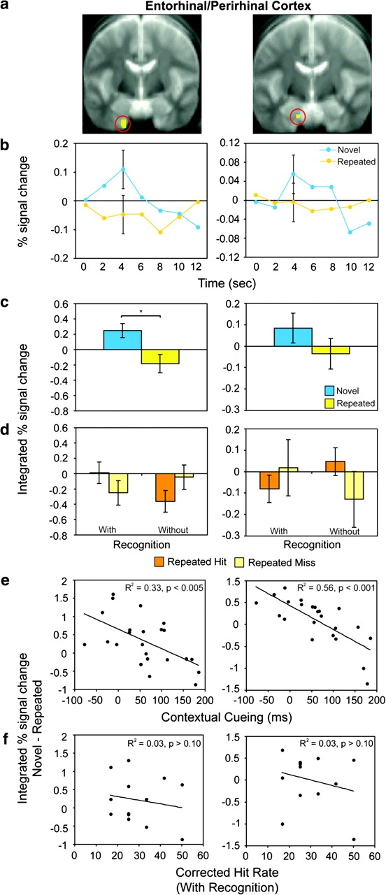
Activation in left entorhinal/perirhinal cortex (−18, −10, −44 and −14, −10, −30) correlated with contextual cueing performance. (a) ROI displayed on group-averaged anatomical images. (b) Time course of activation in each ROI for novel (blue) and repeated (yellow) contexts. (c) Integrated percent signal change in ROI for novel and repeated contexts. (d) Integrated percent signal change for repeated contexts by split by memory performance on the postscan recognition test for both with- and without-recognition memory participants: repeated hit (orange) and repeated miss (light yellow). (e) Integrated percent signal change difference for novel relative to repeated contexts plotted by contextual cueing score, and (f) corrected hit rate for participants with recognition memory. Asterisks indicate significant differences.