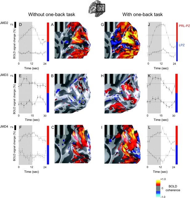
Figure 4.
Viewing the stimuli while performing the OBT elicits a stimulus-synchronized response in the LPZ in JMD2, 3 but not in JMD4. We compare the activation pattern elicited by passive viewing (JMD2, 3) or stimulus unrelated judgments (JMD4) (left panels) and OBT (right panels) of the drifting contrast pattern (insets). The panels from the 3 rows are JMD2, 3, 4, respectively. (A–C) The phase-specified coherence map. (D–F) The average time course during 1 stimulus cycle in the PRL-PZ (top) and the LPZ (bottom). Stimulus-synchronized responses are present in the PRL-PZ and negative or weak responses are present in the occipital pole. (G–L) Stimulus-synchronized responses while performing the OBT, are shown in an identical format to figure (A–F). In JMD2 and 3, a stimulus-synchronized response is elicited in the PRL-PZ and the LPZ, even though there should not be any input from the lesioned retina. In JMD4, a stimulus-synchronized response in the LPZ is not observed, although some regions responded stronger to the mean-luminance presentations (L). Ventral, dorsal, and lateral occipital responses increase during the OBT condition.

