
Figure 3.
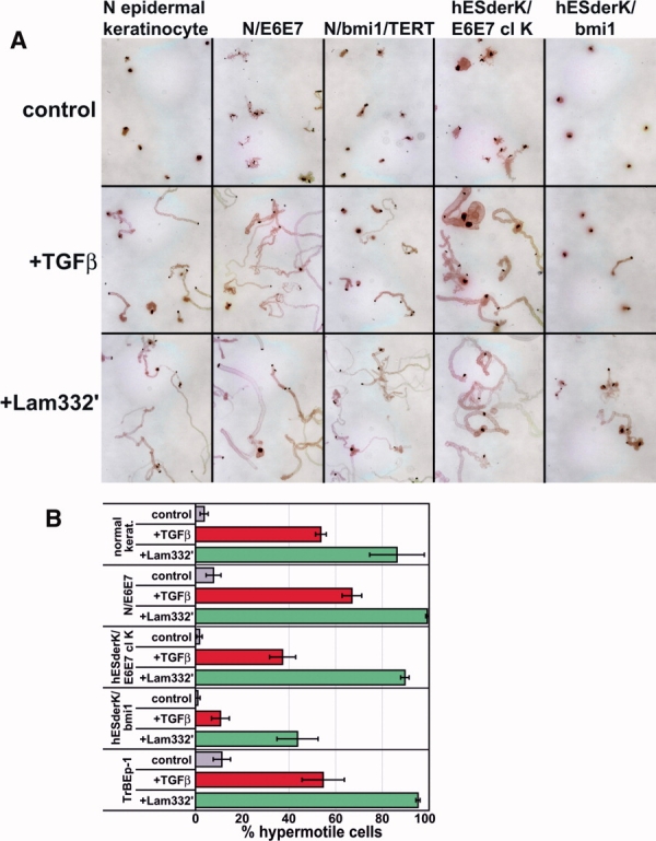
Induction of directional hypermotility in hESderK cells and normal keratinocytes. (A): Directional hypermotility, revealed by immunostaining for Laminin-332, comparing cells 2 days after plating in control conditions, with 0.3 ng/ml TGFβ, and plating on dishes precoated with the γ2 unprocessed form of Laminin-332 (Lam332′). Note the similar, robust motility responses of all cell lines except for hESderK/bmi1, which was responsive but weaker than the other lines. (B): Quantitation of hypermotility responses. Note that a smaller percentage of hESderK/bmi1 cells responded to TGFβ and Lam332′ with hypermotility than did normal epidermal keratinocytes, N/E6E7, and hESderK/E6E7 clone K cells and that normal primary TrBEp-1 were very responsive. Data are the average of three experiments, with error bars showing standard error of the mean. Abbreviations: TGFβ, transforming growth factor beta; TERT, telomerase catalytic subunit.