
Figure 4.
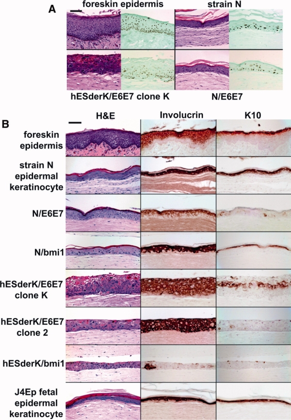
Histogenesis and differentiation-related protein expression by epidermal keratinocyte and hESderK cells in organotypic culture conditions. (A): Histogenesis and p63 expression (left frame hematoxylin and eosin-stained, right frame p63 immunostain with light hematoxylin counterstain), comparing foreskin tissue with organotypic cultures of strain N epidermal keratinocytes, N/E6E7, and hESderK/E6E7 clone K. Bar in top left panel is 100 μm long. (B): Histogenesis and involucrin and K10 expression. Panels show typical fields. Average total epithelial thickness in μm, number of cell layers, and number of stratum corneum cell layers were as follows: for foreskin epidermal tissue (100 μm, 8, 3); organotypic cultures of strain N (60 μm, 5, 2); N/E6E7 (75 μm, 5, 4); N/bmi1 (70 μm, 4, 2); hESderK/E6E7 clone K (140 μm, 10, 0); hESderK/E6E7 clone 2 (75 μm, 5, 0); hESderK/bmi1 (50 μm, 4, 0), and J4Ep (50 μm, 3, 5). Note p63 expression pattern in organotypic cultures similar to that of epidermal tissue in vivo. Note suprabasal expression of involucrin in suprabasal cells of all cell lines tested except for only sporadic expression in hESderK/bmi1 cells, and K10 expression by many suprabasal cells of strain N, N/bmi1, hESderK/E6E7 clone K, and J4Ep but only sporadic expression by N/E6E7, hESderk/E6E7 clone 2, and hESderK/bmi1. Bar in top left panel is 100 μm long.