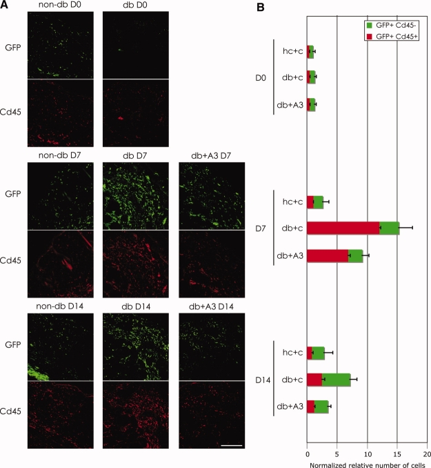
Figure 3.
Expression of HOXA3 in diabetic wounds suppresses inflammatory cell recruitment. (A): Representative images of sections from day 0, 7, and 14 wounds from nondiabetic and diabetic mice treated with control or CMV-HOXA3 plasmid. Upper panels of each group show GFP+ cells and lower panels show Cd45+ cells. Scale bar = 100 μm. (B): Bar graphs showing normalized relative numbers of GFP+ and GFP+Cd45+ (leukocyte) cells recruited to the wounds at days 0, 7, and 14 in nondiabetic and diabetic mice treated with control or CMV-HOXA3 plasmid. Abbreviations: D0, day 0; D7, day 7; D14, day 14; db+A3, diabetic wound treated with CMV-HOXA3; db+A3 D7, diabetic wound treated with CMV-HOXA3 at day 7; db+A3 D14, diabetic wound treated with CMV-HOXA3 at day 14; db+C and db+c, diabetic wound treated with control; db D0, diabetic wound at day 0; db D7, diabetic wound at day 7; db D14, diabetic wound at day 14; GFP, green fluorescent protein; hc+c, heterozygous control + control plasmid; non-db D0, nondiabetic wound at day 0; non-db D7, nondiabetic wound at day 7; non-db D14, nondiabetic wound at day 14.

