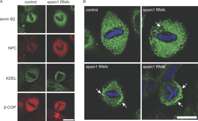
Figure 3.
Mitotic ER network marked by the KDEL antibody is affected by epsin1 reduction in HeLa cells. (A) Confocal images of markers for subcellular compartments in mitosis in control or epsin1 knockdown cells. The maximum projection of images obtained for nuclear lamina (lamin B2), nucleoporins (nuclear pore complex [NPC]; labeled by MAb414), ER (KDEL), and Golgi (β-COP) is shown. (B) Confocal section of mitotic cells stained by the KDEL antibody, which marks the ER network. Arrows indicate areas of uneven ER membrane distribution around condensed chromosomes in mitotic cells after epsin1 knockdown by RNAi, which are not seen in normal control cells. Bars, 10 μm.

