Figure 5.
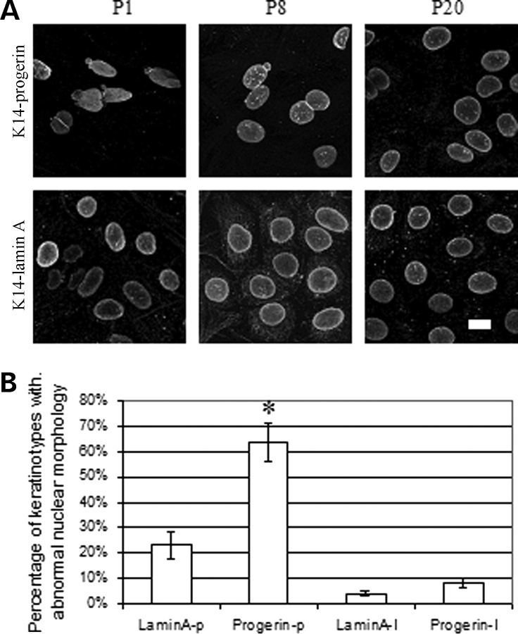
(A) Representative confocal immunofluorescence micrographs of cultured keratinocytes expressing FLAG-progerin (K14-progerin) and FLAG-wild-type human lamin A (K14-lamin A) during spontaneous immortalization by continuous subculture. Top panels show cultured keratinocytes expressing progerin and bottom panels cultured keratinocytes expressing wild-type human lamin A at passage 1 (P1), passage 8 (P8) and passage 20 (P20) labeled with anti-FLAG antibodies. Bar: 10 µm. (B) Percentages of primary cultured mouse keratinocytes at passage 1 expressing wild-type human lamin A (LaminA-p) or progerin (Progerin-p) and spontaneously immortalized keratinocytes at passage 20 expressing wild-type human lamin A (LaminA-I) or progerin (Progerin-I) with abnormal nuclear morphology. Nuclei in 200–500 cells in 25 microscopic fields per sample were scored for abnormal morphology (‘rough’ rim fluorescence, nuclear envelope ‘blebs’) versus normal morphology (smooth, mostly circular nuclear rim fluorescence). Values are means ± SD for n = 5 experiments on different cell lines. *P < 0.05 for progerin-expressing primary keratinocytes at passage 1 (Progerin-p) compared to spontaneously immortalized progerin-expressing keratinocytes at passage 20 (Progerin-I).

