
Figure 7.
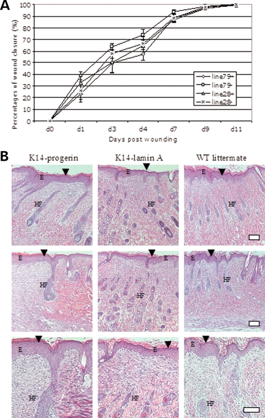
(A) Rates of wound healing in transgenic mice expressing progerin or wild-type human lamin A in epidermis. Graphs show mean percentages of wound closure (y-axis) versus days post wounding (x-axis) for transgenic mice expressing progerin (line 79+), their non-transgenic littermates (line 79−), transgenic mice expressing wild-type human lamin A (line 28+) and their non-transgenic littermates (line 28−). Values are means ± SD for n = 6 wounds in three mice from each group (two wounds per mouse); no differences between groups were significant. (B) Representative sections of skin 11 days after wounding (just prior to complete closure) from transgenic mice expressing progerin (K14-progerin), transgenic mice expressing wild-type human lamin A (K14-lamin A) and a non-transgenic control (WT littermate) stained with hematoxylin and eosin showing no significant differences. Healing skins had thickened epidermis (E) and new hair follicles (HF). Arrowheads show healing wounds. Bar: 50 µm.