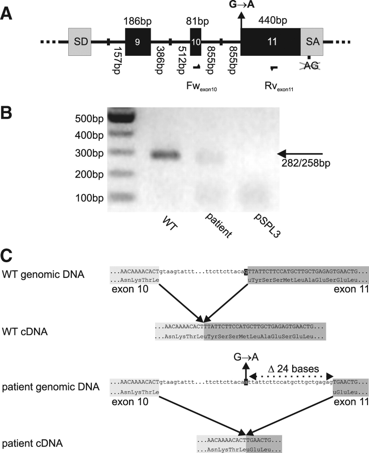
Figure 2:
In vitro splicing of WT and patient minigenes.
(A) Schematic overview of the wild type (WT) and patient minigenes derived from pSPL3. For clarity, only sequence from pSPL3 splice donor (SD) to splice acceptor (SA) is represented. To allow splicing to exon 11, part of pSPL3 SA was deleted. Location of oligonucleotides forward Fwexon10 and reverse Rvexon11 is depicted as arrows. In the patient minigene, mutation G(−1)→A at intron 10–exon 11 boundary is present. (B) RT–PCR on complementary DNA (cDNA) derived from in vitro spliced minigenes using oligonucleotides Fwexon10–Rvexon11. (C) Reconstruction of in vitro splicing using WT and patient minigenes, based on sequencing of in vitro splice products.

