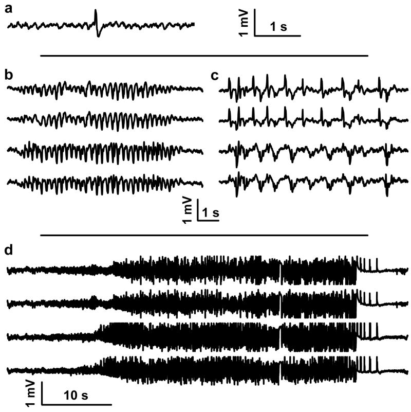
Figure 1. EEG traces recorded from the cortex of rats, 6 days after KA-SE.
(a) Single sharp wave, (b) spike-wave complexes, (c) periodic generalized epileptic discharges, and (d) seizures. Trace in (a) was recorded from a left frontal cortical screw electrode. Traces in (b), (c), and (d) were recorded (top to bottom) from a left frontal, right frontal, left parietal, and right parietal cortical screw electrode.

