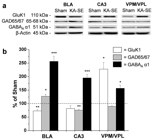
Figure 3. GAD65/67 and GABAA α1 subunit levels are increased in the BLA, while the GluK1 kainate receptor subunit is reduced, on days 7 to 10 after SE.
a Representative Western blots showing GluK1, GAD65/67, GABAA α1, and β-actin antibody binding, in the BLA. For comparison, the levels of these proteins were also measured in the CA3 subfield of the hippocampus, and the ventral posteromedial and ventral posterolateral thalamic nuclei (VPM/VPL). (b) Quantification of GluK1, GAD65/67, and GABAA α1 protein densities relative to β-actin density in KA-SE rats, normalized relative to sham controls. Values are mean ± SEM (n = 6) * p<0.05, **p<0.01 *** p<0.005.

