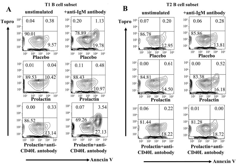
Figure 2. BCR-mediated apoptosis in transitional B cell subsets.
The percentage of apoptotic cells (topro- annexin V+) was determined for T1 and T2 B cell subsets in placebo, prolactin, and prolactin+anti-CD40L antibody-treated mice (n=5 in each group). A) As shown in these representative experiments, after anti-IgM stimulation T1 B cells from mice treated with placebo or prolactin+anti-CD40L antibody showed higher increases in the percentage of apoptotic cells than T1 B cells from prolactin-treated mice (p=0.008 and p=0.003, respectively). B) The degree of anti-IgM-induced apoptosis in T2 B cell subsets of placebo, prolactin and prolactin+anti-CD40L-treated mice was not significantly different.

