
Figure 6.
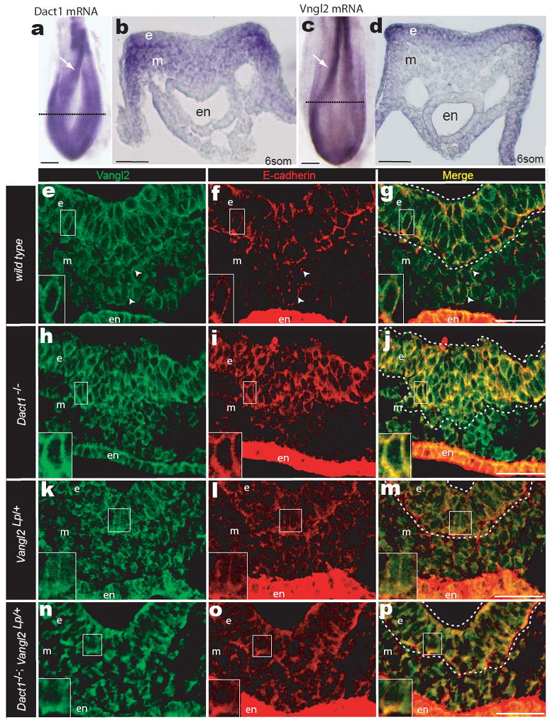
Dact1 and Vangl2 functionally interact at the PS. a, b WISH Dact1 c, d WISH Vangl2 a, c Dorsal aspect showing neural fold (arrow) and approximate level of sections in b & d (dotted lines) b, d Transverse section at PS level. e-p Confocal fluorescent immunohistological localization of Vangl2 (green) and E-cadherin proteins (red) at the PS in Dact1+/+;Vangl2+/+ (wild type; e-g), Dact1-/-;Vangl2+/+ (Dact1 mutant; h-j), Dact1+/+;Vangl2Lp/+ (Loop-tail; k-m), and Dact1-/-;Vangl2Lp/+ (genetically rescued mutant; n-p). Abbreviations as in Figure 2. Scale bars in a-d 0.1 mm, in e-p 0.05mm.