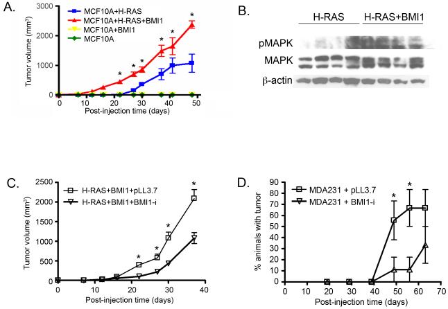
Figure 4. BMI1 expression modulates growth of xenograft tumours.
(A) Mammary fat pad xenografts using MCF10A-derived cell lines. BMI1 and H-RAS overexpression together induce larger tumours over time compared to H-RAS alone. (B) Western blot illustrating activation of MAPK pathway in MCF10A cells overexpressing H-RAS and BMI1. (C,D) Mammary fat pad xenografts using MCF10A+H-RAS+BMI1 (C) and MDA-MB-231 (D) cells with BMI1 knockdown (BMI1-i) or empty vector (pLL3.7). BMI1 knockdown delays tumour formation in MCF10A+H-RAS+BMI1 and MDA231 xenografts (*p < 0.05).

