
Figure 1.
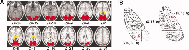
Masks of the thalamus and visual areas and seed thalamic ROIs. (A) Mask covering the thalamus and visual areas (BA 17, 18, and 19). The thalamus and visual areas was extracted using the automated anatomical labeling (AAL) template and the Brodmann template, respectively, in the MRIcro software. Notably, in the Brodmann template, a small part of the vermis was misclassified into the visual areas. This part was manually delineated and excluded from the current mask. The thalamus was displayed in yellow and the visual cortex in red. ‘L’ denotes the left hemisphere of the brain and ‘R’ denotes the right hemisphere. Z‐axial coordinates in the Talairach and Tournoux space are from −24 to 31 mm in steps of 5 mm. (B) The locations of Pulvinar (‘p’ in the figure), Mediodorsal nucleus (‘dm’ in the figure) and Ventrolateral nucleus (‘vl’ in the figure) were shown in a schematic drawing of the thalamus according to the Talairach and Tournoux atlas (Talairach and Tournoux, 1988). The voxels we selected as seed ROI for P, MD, and VL were marked with plus signs.