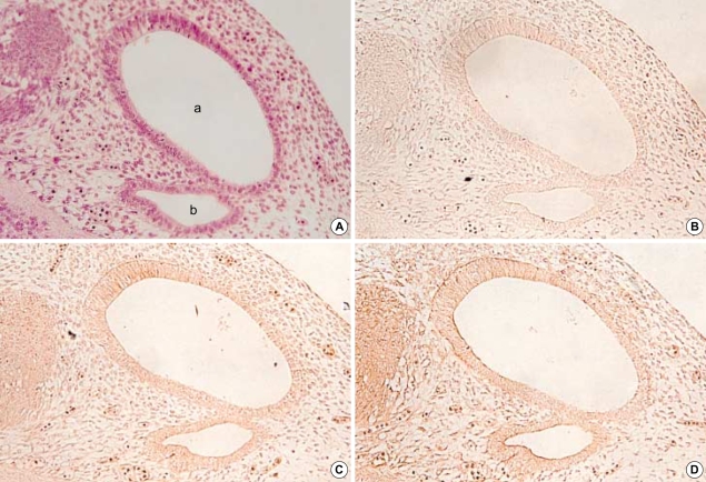
Fig. 1.
Photomicrographs of hematoxylin-eosin stains (A) and immunohistochemical stains using antibodies against TGFβ1 (B), β2 (C), and β3 (D) in the developing internal ear of the 13th-day rat embryo. Horizontal section. The otic vesicle (a) is observed and endolymphatic appendage (b) forms another cavity medial to the otic vesicle. At this stage, the immunoreactivities to all the TGFβ isoforms are very weak (×200).

