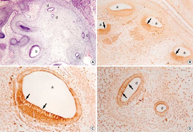
Fig. 3.
Photomicrographs of hematoxylin-eosin stains (A, ×100) and immunohistochemical stains using antibodies against TGFβ2 (B, ×100; C, D, ×400) in the developing internal ear of the 17th-day rat embryo. Sagittal sections. At this stage, cochlear duct (a), ampulla (b), and semicircular canals (c, d) can be identified. In the epithelium of cochlear duct and semicircular canals, strong immunohistochemical reactions of TGFβ2 antibodies are observed, especially at the apices of lining columnar cells (arrows).

