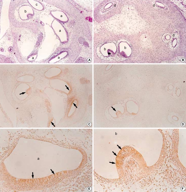
Fig. 5.
Photomicrographs of hematoxylin eosin stains (A, B, ×100) and immunohistochemical stains using antibodies against TGFβ2 (C, D, ×100; E, F, ×400) in the developing internal ear of the 19th-day rat embryo. Sagittal sectons. At this stage, the cochlear duct (a), ampulla (b), saccule (c) and semicircular canals (d) are clearly identified. The immunoreactivity to TGFβ2 was relatively weaker than those of 17th-day embryo is observed at the apices of columnar cells of cochlear duct and crista ampullaris (arrows).

