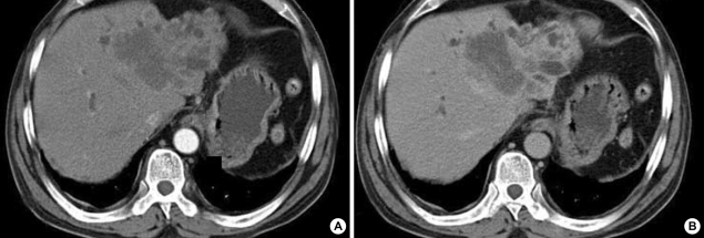
Fig. 1.
Abdominal dynamic CT scan show about 6.5×7×7 cm-sized and ill-defined mass with several daughter nodules in the left lobe. The huge mass with a dilatation of intrahepatic bile ducts is not enhanced on the arterial phase (A), but shows delayed enhancement on the portal phase (B), indicating cholagiocarcinoma.

