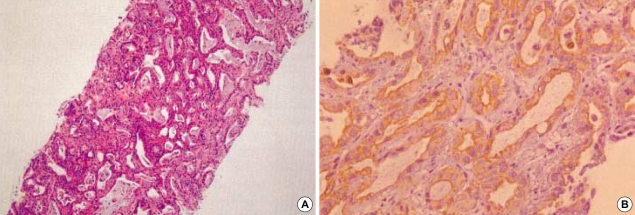
Fig. 2.
Photomicrograph of liver biopsy specimens. Moderately differentiated adenocarcinoma is shown in the hematoxylin-eosin stain (A; original magnification ×100). On the immunohistochemical staining by using cytokeratin 19 (CK 19), dark-brown staining patterns are observed on the epithelium of proliferating bile ducts (B; original magnification ×400).

