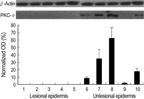
Fig. 2.
Western blot analysis of PKC-α in lesional and non-lesional epidermis of psoriasis patients. Western blotting was performed using mouse anti-PKC-α antibodies, as described in the Materials and Methods. The level of PKC-α expression was quantified by densitometry (optical density, OD) and is expressed as the percentage of the expression of the control β-actin (normalized OD).

