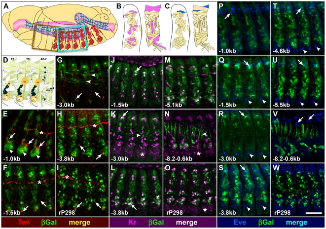
Figure 3. duf enhancer reporters are expressed in specific muscle founder cells.
Cartoon representation of Drosophila embryo depicting different muscles (A) and Kr expression (in B) and Eve expressing DA1 (in C). (D) Brown box region (in A) is enlarged to show Twi expressing adult muscle precursors (AMPs, adapted from [74]). The same region is discussed in (E). (F–W) Confocal projections of stage 14 embryos double labeled with antibodies against β Gal (in green, E–W) and either Twi (in red; E–I), Kr (in magenta; J–O) or Eve (in blue; P–W) corresponding to cyan box region in (A). Wildtype duf expression is seen using rP298 lacZ (I, O and W). Ectopic expression of duf −1.0 kb lacZ (arrowheads in E) is ventral to Twi expressing AMPs (arrows in E). All duf enhancer reporters do not colocalize with Twi in the abdomen (arrows in F–I). Longitudinal visceral muscles are seen in duf −3.0 kb lacZ (arrowheads in G). Non-specific staining of trachea (asterisk in E, F and H). duf −1.5 kb lacZ colocalizes with Kr positive FCs (arrow J). duf −3.0 kb lacZ is expressed in longitudinal visceral muscles (arrowhead) and colocalizes with Kr only in DA1 (arrow in K). duf −3.8 kb lacZ (L) and duf −5.1 kb lacZ (M) colocalize with all Kr positive somatic FCs. duf −8.2−0.6 kb Gal4 expressed in circular visceral muscles (arrowhead in N) do not colocalize with Kr. Kr expression in CNS is marked by asterisk. (P) duf −1.0 kb lacZ is not expressed in DA1 or pericardial cells. duf −1.5 kb, −3.0 kb, −3.8 kb, −4.6 kb and −5.5 kb colocalize with Eve in DA1 (arrows) but not with pericardial cells (Q–U). duf −8.2−0.6 kb Gal4 is not expressed in DA1 (arrows) or pericardial cells (V). Eve expression in the CNS (arrowheads Q–U). Scale Bar = 50 microns.

