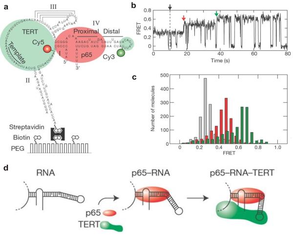
Fig. 4.
Protein-mediated RNA folding in assembly of the telomerase RNP complexes. (a) The Cy3 (donor) and Cy5 (acceptor) fluorophore-labeled full length telomerase RNA molecule immobilized on a surface via a biotin-streptavidin linkage. (b) smFRET time trajectory showing the assembly of the RNP complex after the addition of p65 and TERT (black arrow). The assembly pathway consists of two steps: P65-induced FRET transition (red arrow) and complete assembly of the p65-RNA-TERT ternary complex (green arrow). (c) FRET histogram showing the distribution of the FRET states in the absence of protein (grey), the presence of 10 nM p65 (red) and the presence of both 10 nM p65 and 32 nM TERT1-516 (green). (d) A schematic representation of RNA folding observed in real-time assembly of the telomerase RNP complex. Reprinted by permission from Macmillan Publishers Ltd: [Nature] [84], copyright (2007).

