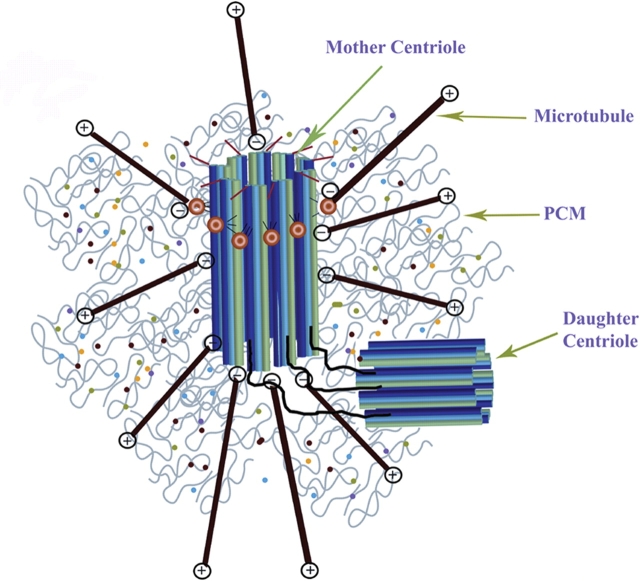
Figure 1.
Schematic diagram of a typical mammalian centrosome composed of two centrioles (mother and daughter centriole) surrounded by a meshwork of pericentriolar material (PCM).
Both centrioles are connected through interconnecting fibers. The mother centriole is distinguished from the daughter centriole by distal and subdistal appendages. The centrosomal material consists of a fibrous scaffolding lattice with a large amount of coiled-coil centrosome proteins and the centrosome's three-dimensional architecture is primarily maintained through specific protein–protein interactions. Microtubules are anchored with their minus ends to the centrosome core structure and microtubule growth is regulated by distal plus-end addition of tubulin subunits.

