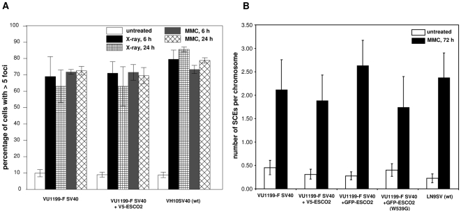
Figure 5. Formation of Rad51 foci and sister chromatid exchanges in RBS cells.
(A) Rad51 foci in normal and RBS cells, as determined 6 and 24 h after treatment with X-ray (12Gy) or MMC treatment (7 µM for 1 h). The percentages of cells containing more than five nuclear foci were determined. Data are the means of at least three experiments; error bars represent the standard error of the mean. (B) SCE induction in RBS cells after MMC treatment. Wild type (LN9SV), RBS (VU1199-F SV40) and RBS cells expressing V5-ESCO2, GFP-ESCO2 or GFP-ESCO2 (W539G) were either mock-treated or treated by continuous exposure to 50 nM MMC. Numbers of SCEs were counted and normalized against the number of chromosomes scored.

