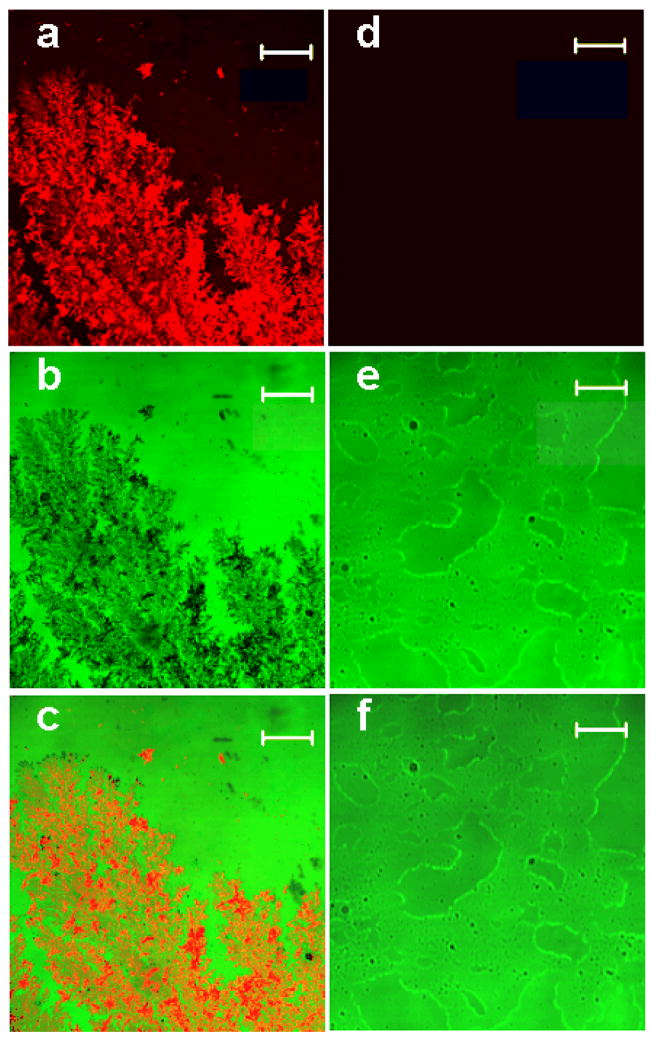
Figure 3.
The laser scanning confocal microscope images. The images of the immunoassay performed on glass slides with fractal-like structures (left column) and without fractal-like structures (right column). (a) The fluorescence images of the immunoassay prepared on the glass slide with fractal-like structures and (d) without the fractals. (b) The transmission images of the immunoassay made on the glass slide with fractal-like structures and (e) without the fractals. (c) The superimposed fluorescence and transmittance images of the immunoassay carried out on the glass slide with fractal-like structures and (f) without the fractals. The bars on all images represent 50 μm.

