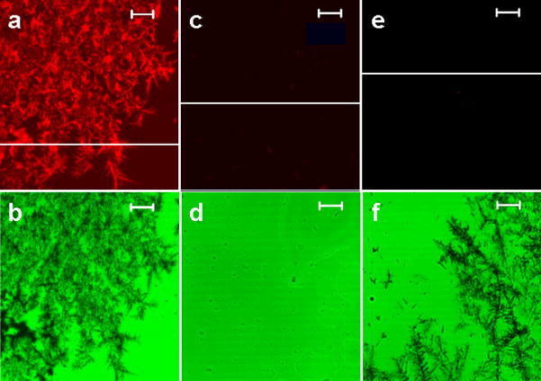
Figure 4.
High magnification laser scanning confocal microscope images. The images of the immunoassay performed on a glass slide with fractal-like structures (left column), the immunoassay performed on a clean glass slide (middle column), and a non-specific binding performed on a glass slide with fractal-like structures (right column). (a) The fluorescence images of the immunoassay prepared on the glass slide with fractal-like structures, (c) the immunoassay prepared on a clean glass slide, and (e) a non-specific binding performed on a glass slide with fractals. (b) The images in transmission mode of the immunoassay prepared on the glass slide with fractals, (d) on a clean glass slide, (f) on a glass slide with fractal-like structures with non-specific protein binding. The bars on all images represent 5 μm.

