Figure 1.
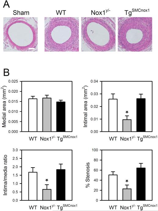
Analysis of neointima formation in WT, Nox1y/- and TgSMCnox1 mice. Injury was induced by insertion of a guide wire into the left femoral artery, and arteries were harvested after 21 days. A. Representative images of arterial cross-sections from WT, Nox1y/- and TgSMCnox1 mice. B. Assessment of medial and intimal area, intima/media ratio, and %stenosis. Areas of media and intima were averaged from duplicate cross-sections, from which intima/media ratio and %stenosis were calculated. Values are means±SE of 13 (WT) and 6 (Nox1y/- and TgSMCnox1) animals. *Significantly different from WT control (p < 0.05). Scale bar=50 μm.

