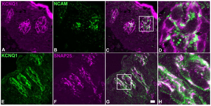
Figure 6.
Single plane confocal images of double immunostaining of human circumvallate sections with antibodies against KCNQ1 and two type III cell markers: NCAM and SNAP-25. Upper panels: a transverse section stained with anti-KCNQ1 (A) and anti-NCAM (B) antibodies; Lower panels: a longitudinal section stained with anti-KCNQ1 (panel E) and anti-SNAP-25 (panel F) antibodies. Overlay of the images (panels C and G) and their high magnification images (panels D and H) showed that all NCAM or SNAP-25 -immunoreactive human taste bud cells displayed KCNQ1 antibody immunoreactivity. Note: To rule out any possible fluorophore effect on imaging, the secondary antibodies conjugated with different fluorophores were used to visualize the KCNQ1 staining on the sections. Scale bar: 20 μm.

