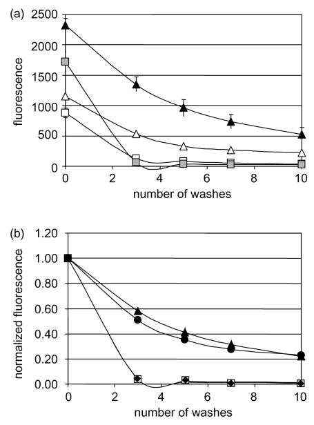
Fig. 3.
CA125/mesothelin-dependant cell adhesion assay. Ninety six-well plates containing OVCAR-3 adherent cells and fluorescently-labeled HEK 293 cells, wild type at 2.5×10⁶ cells/ml (white squares) or 5×10⁶ cells/ml (gray squares); or transfected with mesothelin at 2.5×10⁶ cells/ml (white triangles) or 5×10⁶ cells/ml (black triangles); MPF at 5×10⁶ cells/ml (black diamonds); MSLN1 at 5×10⁶ cells/ml (black circles) were washed up to 10 times. (a) Remaining fluorescence was plotted after each wash or (b) normalized to the original fluorescence for each well. The assay was performed in triplicates and the values were averaged. All standard deviations were less than 4%.

