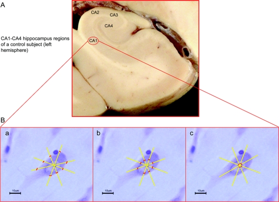
Figure 1 Cell body, nuclear, and nucleolar markers for morphometric measurements
(A) Large coronal section of a formalin-fixed brain of a control subject (left hemisphere). (B) Single pyramidal neuron from the CA1-hippocampus region, the specific markers for the morphometric measurements of cell body (a), nucleus (b), and nucleolus (c). The pictures of a, b, and c were realized with a digital camera connected with a light microscope using an oil-objective at 100× magnification.

