
FIG. 1.
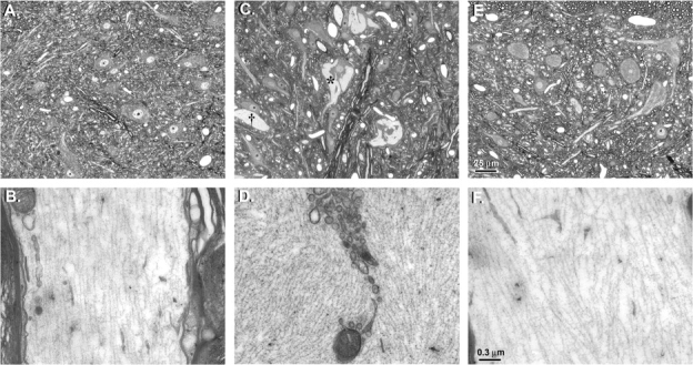
Cross-sections of lumbar spinal cords of rats treated i.p. with vehicle (A and B), 20 mg/kg/day 1,2-DAB (C and D), or 1,3-DAB (E and F), 5 days a week, for 2 weeks. The upper panel shows light-microscope images and the lower panel are the corresponding electron microscopy images. Hypochromic zones (*) corresponding to NF accumulation in anterior horn cells and giant axonal swellings (†) were evident in animals treated with 1,2-DAB (C). Electron microscopy of this region revealed disorganization of the cytoskeleton and clustering of organelles in axonal swellings that were densely packed with NF (D). Spinal cords from animals treated with vehicle (A, B) or 1,3-DAB (E, F) were unremarkable. Cross-sections (∼900 nm) for light microscopy were stained with 1% toluidine blue. Thin sections (∼90 nm) of regions of interest were stained with 2% uranyl acetate followed by 1% lead citrate for transmission electron microscopy.