Figure 1.
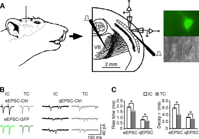
Intracortical and thalamocortical synapses possess different transmission kinetics. A, Left and middle, Schematic drawing of the in vivo injection of viral constructs, in vitro stimulating and recording electrode locations in the thalamocortical slice preparation. Hip, Hippocampus; VB, ventral basal nucleus. Right, Simultaneous recordings, made under transmitted light illumination (bottom), from pairs of recombinant protein-expressing neurons, identified by GFP fluorescence (top), and nearby nonexpressing control neurons in layer 4. B, Left traces, evoked EPSCs (eEPSCs) from intracortical (IC) and thalamocortical (TC) synapses from a pair of layer 4 neurons. Note the paired-pulse facilitation of intracortical responses and depression of thalamocortical responses of both control nonexpressing and expressing neurons. Right traces, Asynchronous quantal EPSCs (qEPSCs) from intracortical and thalamocortical synapses in a layer 4 neuron. C, Left, Rise time of eEPSPs (IC, 1.87 ± 0.12 ms; TC, 1.52 ± 0.12 ms; n = 18; p < 0.05) and qEPSCs (IC, 0.78 ± 0.03 ms; TC, 0.63 ± 0.03 ms; n = 20; p < 0.005) from intracortical and thalamocortical synapses. Right, Decay time constant of eEPSPs (τ; IC, 5.70 ± 0.47 ms; TC, 3.91 ± 0.32 ms; n = 18; p < 0.005) and qEPSCs (τ; IC, 3.16 ± 0.14 ms; TC, 2.49 ± 0.11 ms; n = 20; p < 0.0005) from intracortical and thalamocortical synapses. *p < 0.05 (Wilcoxon's test).

