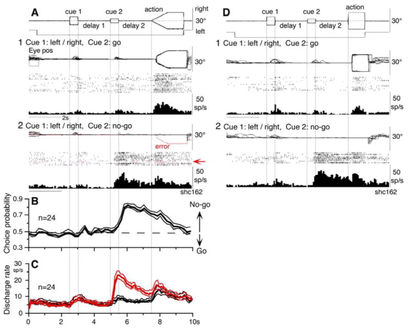
Fig. 6.

No-go neurons. A and D, representative neuron during smooth pursuit (A) and saccade tasks (D). A1, go trials when cue 1 was rightward and leftward visual motions. A2, no-go trials. Red traces (arrows) highlight an error trial. B, CP time course for 24 no-go neurons during no-go and go- trials. C, time course of mean (±SE) discharge of the 24 neurons during no-go (red) and go-(black) trials. D1 and D2, go trials and no-go trials for saccades, respectively.
