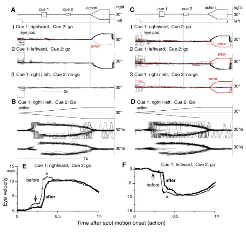
Fig. 8.

Inactivation of bilateral SEF. Eye position (A, C) and velocity (B, E, D, F) aligned at the onset of cue 1 before muscimol infusion (A, B) and after infusion (C, D). E and F compare de-saccaded and averaged eye velocity before (thin lines) and after (thick lines) infusion for rightward (E) and leftward pursuit (F) correct performance. De-saccaded portions were connected by straight lines. See text.
