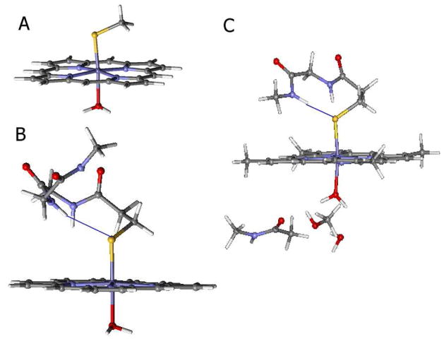
Figure 2.
Structural models for the calculations of the resting ferric site used in this study. A) Porphyrin ligand with water and thiolate axial ligands; B) An expanded to include H-bonding from the backbone to the thiolate (indicated by a blue line); and C) B expanded to include H-bonding to the ligand in the distal pocket. For the substrate-bound site, the H2O was eliminated and models A and B applied.

