
Figure 1.
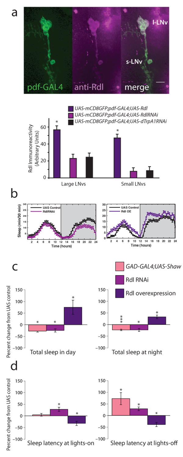
LNvs express the Rdl GABAA receptor and mediate GABAergic effects on sleep. a, Top shows images of Rdl expression in wild type LNvs. Adult brains from pdf-GAL4;UAS-mCD8-GFP animals were stained with anti-Rdl (1:100) and visualized with confocal microscopy. Rdl is shown in magenta, GFP in green, overlap in white. Scale bar = 10 μm. Bottom shows quantification of somatic Rdl levels in LNvs expressing excess Rdl, RdlRNAi or control dTrpA1RNAi. b, Standard sleep plots of control and experimental flies in 12 hour: 12 hour light:dark (LD). Left panel shows the effects of reducing Rdl levels in LNvs: pdf-GAL4;UAS-RdlRNAi, right panel shows the effects of overexpressing Rdl in LNvs: pdf-GAL4;UAS-Rdl. c, GABA regulates total sleep. 12 h sleep from the light (left) or dark (right) period in LD was assessed for animals with decreased overall GABAergic transmission (GAD-GAL4;UAS-Shaw; n=62), decreased LNv Rdl levels (pdf-GAL4;UAS-RdlRNAi; n = 93), or increased LNv Rdl levels (pdf-GAL4;UAS-Rdl; n = 21). Data are expressed as the percent change from the genetic control. d, GABA regulates sleep onset. The latency to first sleep bout during the light (left) or dark (right) period in LD was assessed for the same genotypes. Data are expressed as the percent change from the genetic control. * indicates P < 0.05, ** P < 0.005 and *** P < 0.0005 for comparisons of experimental and control using ANOVA with Tukey posthoc test.