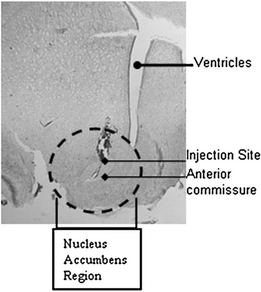
Fig. 2.
Nissl-stained hemisection in the coronal plane shows an example of the site of injection. HSV vectors were injected into the NAc as described in the Experimental procedures. Stereotaxic coordinates were determined from the Rat Atlas (Paxinos and Watson, 2002). The section is at A/V +1.5, M/L±1.6 and D/V−7.6 and 1.6×1000 magnification. The anterior commissure, ventricles and injection site are delineated and the accumbens region has been circled to orient the reader.

