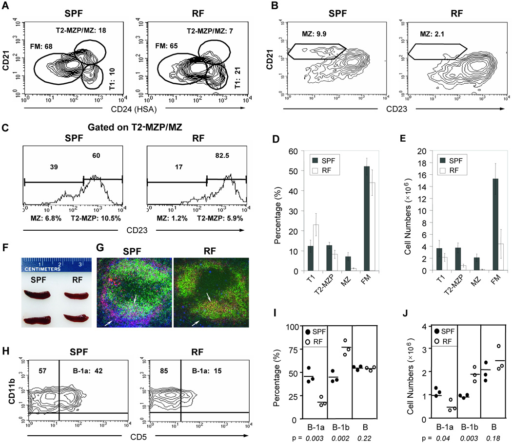
Figure 1. Mice bearing distinct resident microbiota (RF mice) exhibit a severe reduction in MZ and B-1a B cells.
(A) CD21/HSA(CD24) and (B) CD21/CD23 profiles of splenic CD19+ B cells from C57BL6 mice maintained in SPF versus RF conditions. The relative percentages of transitional 1 (T1), transitional 2 marginal zone precursor (T2-MZP) + marginal zone (MZ) (T2-MZP/MZ), follicular mature (FM), and marginal zone (MZ) are indicated. (C) CD21/CD23 profiles, gated on the T2-MZP/MZ (CD21hiHSAhi) population in (A) to distinguish (CD23hi) MZP from (CD23lo) MZ cells. (D) Percentage and (E) absolute numbers of T1, T2-MZP, MZ, and FM splenic B cell populations, with averages from five age-matched pairs. The relative percentage of MZP and MZ B cells were derived by multiplying the CD23hi or CD23lo fractions in (C) with the T2-MZP/MZ fractions in (A). (F) Comparison of spleens from age and gender-matched SPF and RF mice. (G) Immunofluorescence staining of splenic tissue sections with IgD (green), IgM (red) and MOMA-1 (blue) against marginal metallophilic macrophages. The arrows indicate marginal zone B cells, based on IgMhigh staining (red) and localization outside the MOMA+ layer (blue). Arrowheads highlight B cells in follicular areas, with IgD and IgM double expression confirmed by yellow staining. (H) Peritoneal cells (PECs) were gated on B220, and the staining contours for CD11b (Mac1) and CD5 are shown for SPF and RF mice. Minimal B-2 B cells (B220-gated, CD11b−CD5−) were detectable in this compartment. (I) Percentage and (J) absolute numbers of B-1 subsets in SPF (solid circles) and RF (open circles) animals. Representative results of 3 or more similar experiments.

