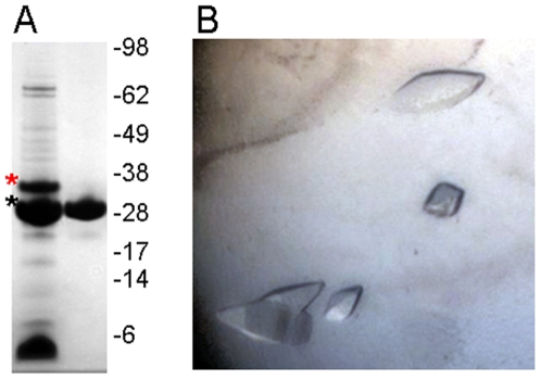
Figure 2. Purification and crystallization of paraplegin305–565.
A. Coomassie-stained SDS-polyacrylamide gel showing the purity of crude paraplegin305–565 after TEV-cleavage (left lane; red asterisk, hexahistidine-tagged protein; black asterisk, cleaved protein), and after the final purification step (right lane). B. Example of crystals grown under the conditions that yielded diffraction data.

