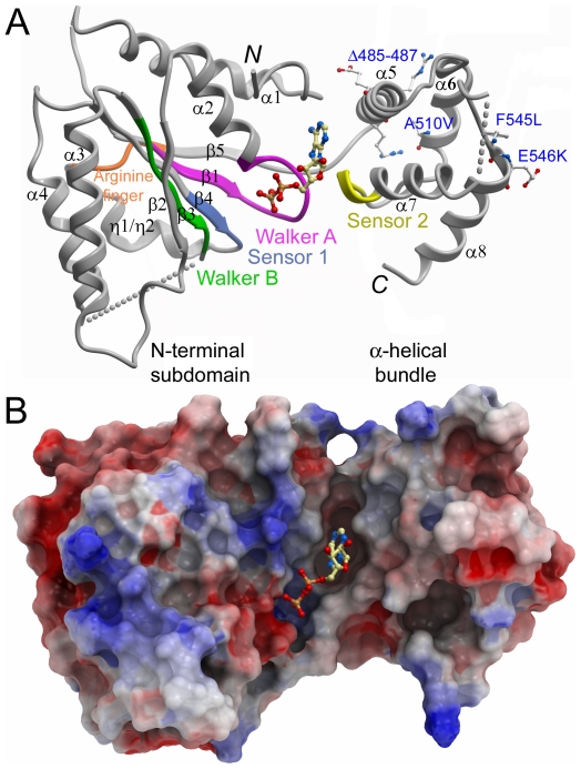
Figure 3. Overview of the paraplegin ATPase domain structure.
A. Schematic representation of the crystal structure of a monomer of paraplegin305–565 with bound ADP. Sequence motifs indicated in the sequence alignment in Figure 1 have been mapped onto the structure. The positions of disease-related residues are labeled in blue. B. Electrostatic surface representation of paraplegin305–565 illustrating the nucleotide binding cleft.

