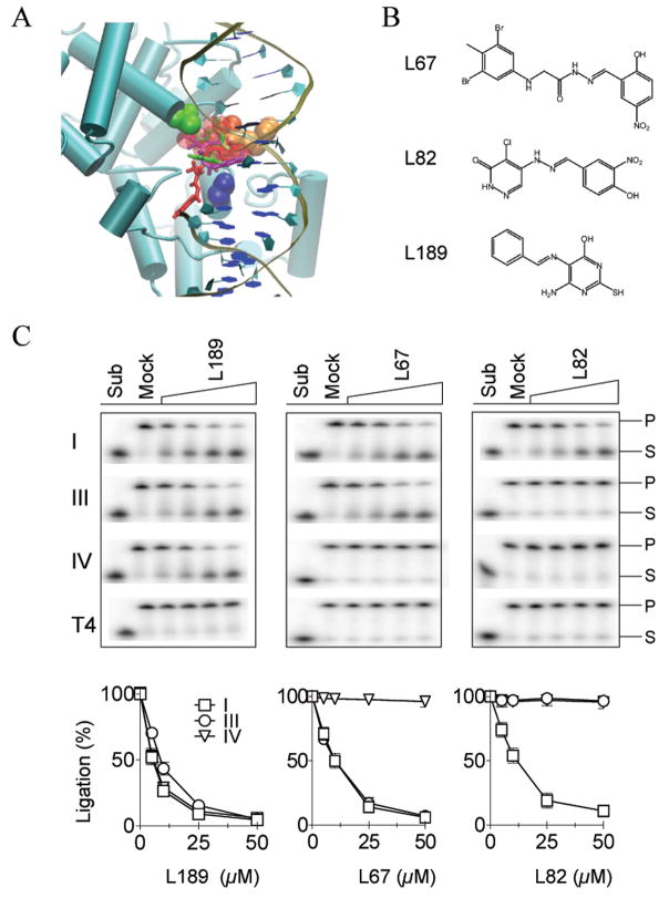
Figure 1. Small molecule inhibitors of human DNA ligases identified by CADD.
A Key residues in the DNA binding pocket, Gly448 (green) Arg451 (orange) and Ala455 (blue), within the hLigI DBD (aqua ribbon format) are shown in VDW representation with the nicked DNA in cartoon format. The sphere set used to direct the docking of small molecules is indicated by red transparent spheres. Docked orientations of the three characterized compounds, L67 (purple), L82 (red), and L189 (green). B. Chemical structures of L67, L82 and L189. C. Representative gels of DNA ligation assays. The results of three independent experiments are shown graphically. For clarity, the data for T4 DNA ligase, which was not significantly inhibited, has been omitted (hLigI, □; hLigIIIβ, ○; hLigIV/XRCC4, ▽).

