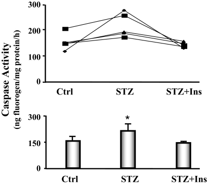
Figure 6.
Caspase-3 activity is increased in the cardiac muscle of diabetic mice. The activity of caspase-3 in cardiac muscle was measured using a fluorogenic substrate (Ac-DEVD-amc), in the presence or absence of the caspase-3 inhibitor (Ac-DEVD-CHO). Five feeding-matched triplets of mice were monitored. Each triplet contained one mouse with diabetes (STZ), one pair-fed, sham-injected control (Ctrl), and one pair-fed diabetic mouse receiving insulin treatment (STZ+Ins). Triplets are connected by a line (line graph). The bar graph shows the average caspase-3 activities. Data are reported as the means ± se (n = 5 per group). *, P < 0.05 vs. control.

