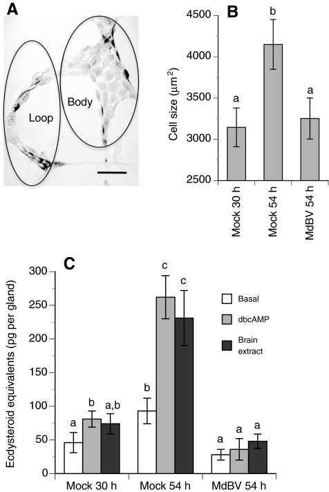
Fig. 6.
Prothoracic gland morphology and in vitro release of ecdysteroids (means ± s.e.) by glands from MdBV- or mock-infected P. includens fifth instars. (A) Light micrograph of a prothoracic gland from a 30 h fifth instar showing two domains designated as the loop and gland body region. Scale bar, 100 μm. (B) Estimated size of prothoracic gland cells. Fifth instars were injected with MdBV or Grace's medium only (Mock) at 30 h. Cell size was then determined for prothoracic glands from mock-infected larvae immediately after injection of medium only (30 h) or 24 h later (54 h). Cell size for prothoracic glands from MdBV-infected larvae was determined 24 h after infection (54 h). Cells from three prothoracic glands were measured per treatment. Bars with different letters are significantly different from one another (Tukey–Kramer HSD, P≤0.05). (C) In vitro release of ecdysteroids (means ± s.e.) by prothoracic glands from MdBV- or mock-infected fifth instars. Prothoracic glands were collected immediately after injection (30 h) or 24 h post-injection (54 h) as described in B. Basal, dbcAMP-stimulated and brain extract-stimulated levels of ecdysteroid released by glands were then determined. A minimum of four prothoracic glands were bioassayed per treatment and time point. Bars with different letters are significantly different from one another (Tukey–Kramer HSD, P≤0.05).

