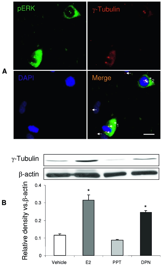
Figure 8.
E2-induced ERK phosphorylation promotes centrosomes amplification. A. pERK immunoreactivity was observed in the dividing cell nuclear and cytosol located centrosome as demonstrated by double immunostaining of pERK (green) and centrosome marker, γ-tubulin (red). Basic nucleotides were counterstained with DAPI (blue). Dividing cells (*) are pERK positive and display two centrosomes (orange, indicated by arrowheads), indicating the colocalization of pERK (green) and γ-tubulin (red). In contrast, nondividing cells are pERK negative and exhibit only one centrosome (red, labeled only by γ-tubulin, white arrows). hNPC*1 hNPC, two centrosomes are located on both sides of the metaphase plate indicating entry into metaphase. hNPC*2 cell exhibited two centrosomes located to one side of the condensed chromosome indicating that this cell is likely in prophase. B, The centrosome marker protein, γ-tubulin, expression was detected in hNPCs by Western blot. Both E2 and DPN increased γ-tubulin levels in hNPCs, whereas PPT did not. Activation of ERβ either by E2 or DPN increased γ-tubulin protein levels in hNPCs and is indicative of amplification of centrosomes. Loading control, β-actin, normalized data are presented as mean ± sem from three independent experiments. *, P < 0.05 vs. control or as indicated.

