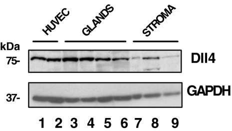
Figure 2.
Western blot analysis of Dll4 in HUVECs and in human endometrial cells. Total protein (100 μg) from cell lysates was applied to each lane: HUVECs (lanes 1 and 2), glandular cells (proliferative, early, and two late secretory phase, lanes 3–6), and stromal cells (proliferative and mid and late secretory phase, lanes 7–9, respectively). The upper and lower panels show the intensity of Dll4 and GAPDH bands, respectively. The relative intensities of Dll4 bands, normalized by GADPH, were 1.1 (HUVEC); 2.7, 1.7, 0.9, and 0.6 (glandular cells); and 0.3, 1.0, and 0.4 (stromal cells), respectively.

