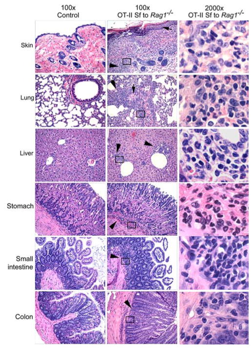
FIGURE 5.
Vβ5neg T cells from OT-II Sf mice transferred multiorgan inflammation in Rag1−/− recipients. Vβ5neg T cells were prepared from OT-II Sf mice as described in Materials and Methods. Rag1−/− recipients (n = 3, 6 wk old) were transferred (i.v.) with 6 × 10⁶ cells of the purified population. Inflammation in various organs/tissues was determined 12 wk after transfer by examination under a microscope of H&E-stained tissue sections (middle column; original magnification, ×100; right column; original magnification, ×2000). Untreated Rag1−/− mice were used as control (left column; original magnification, ×100). The right column panels demonstrate the presence of mononuclear and polymorphonuclear cells in the boxes in the middle column. The arrows indicate thickened skin and bronchial epithelium. The arrowheads indicate regions of leukocytic infiltration.

