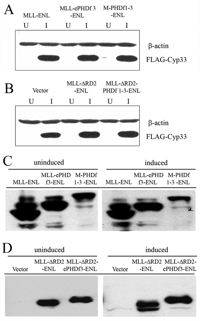
Figure 3. Expression of Flag-Cyp33 and fusion proteins in tetracycline treated or untreated 293 cells.
Western blot analysis was performed on enriched CD2+ 293 cells transfected with fusion protein constructs. The induced Cyp33 (A and B) or the fusion protein (C and D), were detected using anti-flag antibody. β-actin was used as a loading control. U, uninduced; I, induced

